CT / USG guided RFA and Biopsy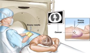
Needle biopsy is a medical test performed by interventional radiologists to identify the cause of a lump or mass, or other abnormal condition in the body. During the procedure, the doctor inserts a small needle, guided by X-ray or other imaging techniques, into the abnormal area. A sample of tissue is removed and given to a pathologist who looks at it under a microscope to determine what the abnormality is — for example, cancer, a noncancerous tumor, infection, or scar.
Biopsy/FNAC of lesions can be done depending on their locations.
4D Color Ultrasound ( Foetal – Renal – Carotid – Peripheral Vessels )
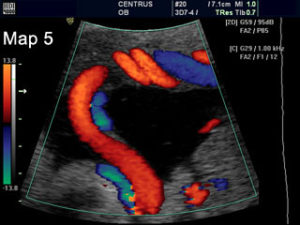
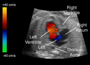 A Doppler ultrasound, also called a Color Doppler test is a non-invasive test that can be used to estimate your blood flow through blood vessels. It helps doctors evaluate blood flow through major arteries and veins, such as those of the arms, legs, and neck. It can show blocked or reduced flow of blood through narrow areas in the major arteries of the neck that could cause a stroke. It also can reveal blood clots in leg veins (deep vein thrombosis, or DVT) that could break loose and block blood flow to the lungs (pulmonary embolism). During pregnancy, Doppler ultrasound may be used to look at blood flow in an unborn baby (foetus) to check the health of the foetus.
List of Common Color Doppler test procedures:
• Abdominal
• Carotid
• Gravid Uterus
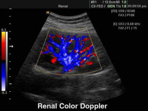
• Renal
• Single Limb Doppler (Arterial and Venous)
• Dual Limb Doppler (Arterial and Venous)
A renal ultrasound is a safe and painless test that uses sound waves to make images of the kidneys, ureters, and bladder. The kidneys are a pair of bean-shaped organs located toward the back of the abdominal cavity, just above the waist. They remove waste products from the blood and produce urine.
Transvaginal ultrasound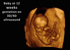
Transvaginal ultrasound uses a probe that is inserted directly into the vagina. It is performed in the doctor’s office similar to a pelvic examination. This type of exam is most commonly used in the early weeks of pregnancy to rule out suspected problems or to assess the gestational age of the embryo. In early pregnancy this examination can provide more accurate information then a transabdominal examination.
High-Resolution Ultrasound Musculoskeleta
High-resolution ultrasound is an extremely useful and versatile technique for examining the musculoskeletal system. It has the advantage of being readily available and inexpensive, at the same time providing highly detailed information regarding a variety of clinically relevant structures of the musculoskeletal system, including tendons, muscles, nerves, ligaments, joints and bone.
 Vikas Diagnostics Best Diagnostics Lab In Kanpur

